

After an HIE diagnosis, families often find themselves learning an entirely new language:
MRI. EEG. Sarnat score. Seizure burden. Basal ganglia. Watershed injury. Background activity.
And behind almost every new term is the same question: “Does this tell us what our child's future will look like?”
These tools can give clinicians important information, but prognosis after HIE works best when we think in terms of probability rather than certainty.
HIE neuroprognostication usually involves combining multiple pieces of information.
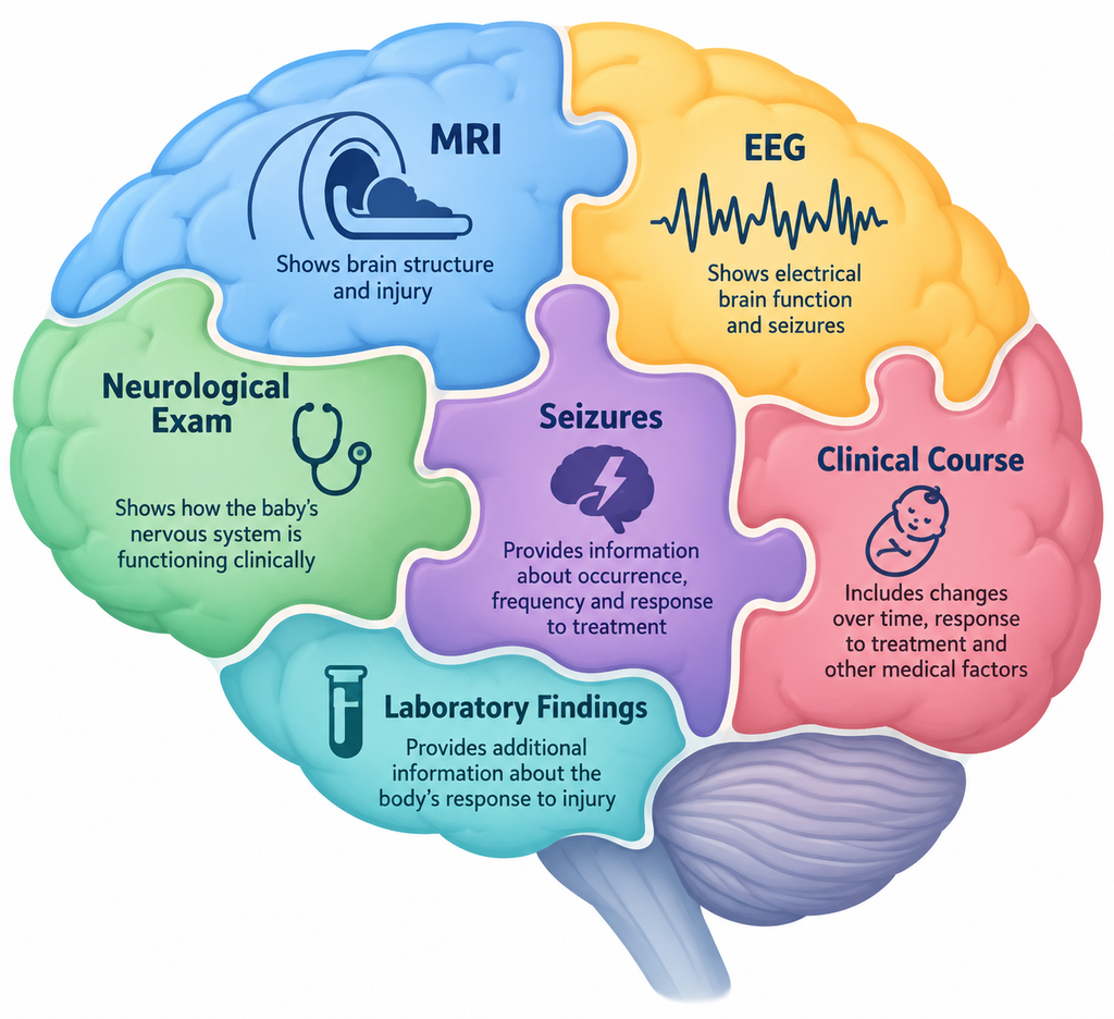
These can include:
Each adds information. None independently tells us everything, because we know some things are modifiable with therapy and other interventions, supports and services.
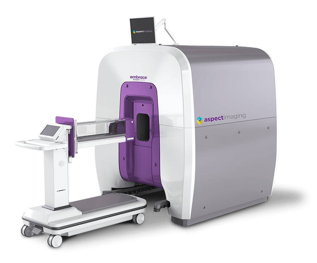
Magnetic resonance imaging, or MRI, is a type of brain imaging that uses a powerful magnetic field and radio waves to create detailed pictures of the brain. It does not use radiation. With HIE, MRI is used to look for evidence of brain injury, including where injury occurred, how extensive it may be, and what areas of the brain were affected.
MRI is one of the most valuable tools available for understanding brain injury after neonatal HIE.
Different patterns of injury can provide important information. For example, injury involving deep gray structures such as the basal ganglia and thalamus may carry different developmental risks than predominantly watershed-pattern injury involving other regions of the brain.
The location, pattern and extent of injury can therefore help clinicians estimate the likelihood of different developmental outcomes.
An MRI is not a photograph of a child's future. It is a snapshot in time.
MRI findings need to be interpreted alongside the baby's neurological examination, EEG, seizures, clinical course and other information. Timing also matters, because HIE-related changes evolve after the injury and what is visible on imaging can depend partly on when the MRI is performed.
Most importantly, MRI cannot directly tell us how an individual child will eventually learn, communicate, move, regulate emotions, build relationships or function in a classroom.
It provides important information about risk. It does not provide certainty about an individual child's developmental future.
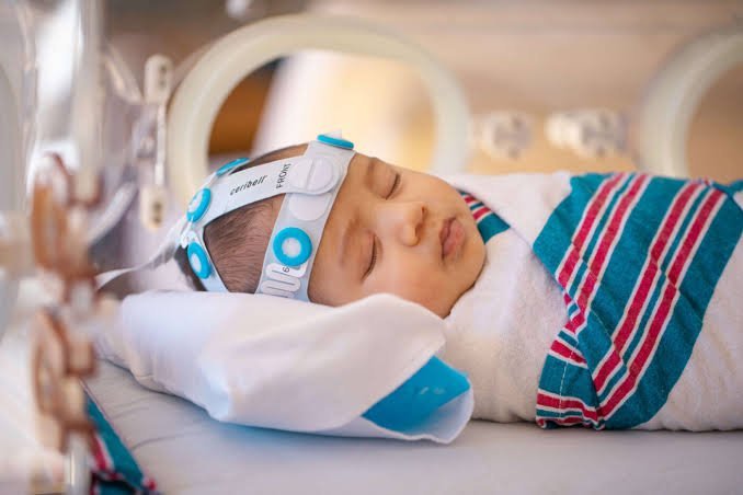
An electroencephalogram, or EEG, records the brain's electrical activity using small electrodes placed on the scalp. In babies with HIE, EEG monitoring is especially important because neonatal seizures are common and many seizures in newborns have few — or no — outward signs.
Continuous EEG can help the medical team identify seizures, understand how frequently they are occurring, determine how long they last, and evaluate whether treatment is working.
But EEG provides more information than seizures alone.
Clinicians also evaluate the baby's background brain activity — the ongoing electrical patterns occurring between seizures. Features of the EEG background, including how organized or suppressed it is and how it changes over time, can provide information about the severity and evolution of encephalopathy.
That makes EEG both a diagnostic and monitoring tool and one piece of the larger prognostic picture.
Research in mild HIE illustrates this well. In a cohort followed to age five, Murray and colleagues examined early EEG findings alongside later neurodevelopmental outcomes.
Early EEG grade was associated with outcome, but the study also demonstrated something important about long-term follow-up: children classified as having mild HIE still showed group-level differences in cognitive outcomes at age five compared with controls.
The neonatal EEG provided valuable information.The developmental follow-up was still necessary.
Like MRI, EEG helps clinicians understand risk and brain function at that point in time. It cannot independently predict everything about a child's future development.
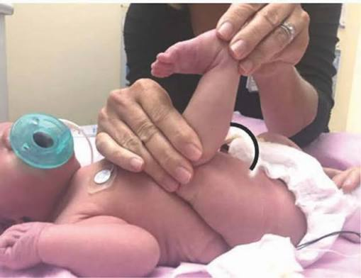
A neonatal neurological examination is a structured assessment of how a baby's nervous system is functioning. Unlike MRI, which shows brain structure, or EEG, which measures electrical activity, the neurological exam allows clinicians to observe how the baby's brain and nervous system are functioning clinically.
The Sarnat examination and related standardized neurological assessments have long been used to help classify neonatal encephalopathy as mild, moderate or severe.
This classification has been especially important because the original therapeutic hypothermia trials used clinical evidence of moderate-to-severe encephalopathy, together with other eligibility criteria, to identify babies for cooling treatment.
For babies with suspected HIE, clinicians assess areas such as:
The neurological examination also isn't necessarily a one-time event.
A baby's examination can evolve during the first hours and days after birth. Medication, seizures, illness, therapeutic hypothermia and other clinical factors can influence what clinicians observe.
For that reason, repeated or serial neurological examinations can provide valuable information about whether encephalopathy is improving, remaining stable or evolving.
But the examination has limitations too.
A newborn neurological exam cannot assess future reading ability, executive functioning, attention, memory, complex language, school performance or many of the other higher-order abilities that develop over childhood.
It tells clinicians something very important: How is this baby's nervous system functioning right now?
It does not, and cannot, answer every question about how that child's nervous system will function years from now.
MRI shows us important information about brain structure and injury.
EEG shows us important information about electrical brain function and seizures.
The neurological examination shows us important information about how the baby's nervous system is functioning clinically.
Each gives us a different window into the brain.
That's why the best HIE prognostication doesn't hinge on one MRI finding, one EEG pattern, one Sarnat score, or any other single result.
Clinicians bring these pieces together alongside seizures, the baby's clinical course, laboratory findings and how the baby changes over time.
Even then, the result is usually: “This increases or decreases the likelihood of…” rather than: “This guarantees…”
That distinction matters.
When discussing prognosis, consider asking:
Those questions turn prognosis into a conversation rather than a verdict.
MRI matters. EEG matters.The neurological examination matters.
But your child is not an MRI, an EEG or a Sarnat score.
These tools help clinicians estimate risk. They cannot fully predict an individual child's developmental path. The most responsible approach combines the best available evidence with honest uncertainty and meaningful long-term follow-up.
Visit our HIE Outcomes resource for more information about prognosis, outcome research and the range of developmental paths after HIE.
Connect with families, read inspiring stories, and get helpful resources delivered right to your inbox.


Connect with families, read inspiring stories, and get helpful resources delivered right to your inbox.


